
Pelvic Ultrasound Can Keep You Health
The pelvic ultrasound procedure generally is painless and safe. Your doctor uses sound waves to take a look at certain images of your pelvis. With this examination, the ultrasound machine will send sound waves into your pelvic, displaying images that get recorded on a computer. The images are black and white, showing the inner structures of your pelvis. Images such as your bladder, as well as with girls, their fallopian tubes, cervix, uterus and ovaries are shown.

Why may you need a pelvic ultrasound?
Typically, your doctor can order a pelvic ultrasound if he/she may be concerned that there are some problems in your pelvis. The doctor can look at or measure your organs of the pelvis with an ultrasound.

They can assess the following:
Where the ovaries and uterus are, as well as for their shape and size
To see the density and thickness of organs and tissues that are in the pelvis
Masses or fluids that are in your bladder, fallopian tubes, the uterus’s muscles or the endometrial
The thickness and length of the cervix
Any shape changes of the bladder
To look at the flow of blood through the organs of the pelvic
This procedure can give your doctor enough information about all the above-mentioned problems, but it’s not possible to provide exact identification for a particular disease such as cancer.
A pelvic ultrasound can also help your doctor with the following:
To discover any problems that are in your uterus’s structure, plus assessing the condition of the endometrial.
Your doctor can find in your pelvis cysts, masses, fibroid or any other type of tumor.
They can find your intrauterine contraceptive device or IUD.
The doctor will be able to diagnose a pelvic inflammatory condition or any other type of infection or inflammation.
After your menopause, if you experience bleeding, the doctor can discover the cause.
If you get treatment because of infertility, they can monitor your ovaries.
In the case of Vitro fertilization, he/she can collect eggs and fluid from your follicles/egg sacs.
It will be possible to diagnose an ectopic pregnancy that ordinarily forms in your fallopian tube on the outside of your uterus.
During your pregnancy, they can monitor how the fetus grow.
The doctor can also glance at specific fetal conditions.
It’s also possible to use the ultrasound in procedures like endometrial biopsies, or a transvaginal ultrasound that is used in a Sono hysterography procedure. With the transvaginal, the uterus will be filled with a fluid to give the doctor an enhanced image.
Information about a pelvic ultrasound
It’s a diagnostic examination, it is a noninvasive procedure and it produces some images to evaluate structures and organs within your pelvis. It includes your ovaries, fallopian tubes, vagina, cervix, and uterus.
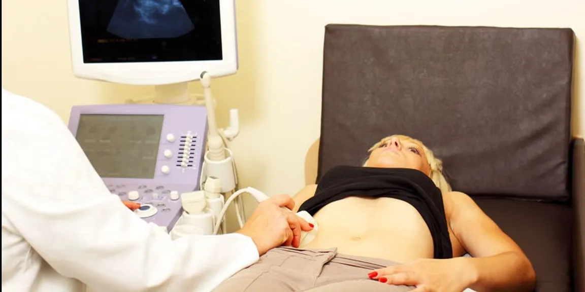
The ultrasound makes use of a transducer, which will send ultrasound waves out at such a high frequency that it won’t be heard. This transducer gets placed on your skin, with the sound waves moving through your body to all the structures and organs inside. These waves bounced off your organs the same as an echo, after which it returns back to the transducer. Which will process this reflection of waves, and the computer will convert it into the image of your tissues or organs that’s been examined.
These waves can travel at speeds that vary from each other, depending usually on what kind of tissues it encounters. Through your bone tissues it can go the fastest, and through the air, it will be the slowest. With the speed that these waves return and also the amount that return, establishes the different tissue types when the transducer translates it.
The doctor will apply a gel onto your skin and then the transducer. This allows for an even movement over your skin with the transducer, as well as to eliminate any air that can come in between the transducer and your skin. This is done in order to provide the best conduction of sound.
The doctor can use either one or sometimes both of the following two methods for a pelvic ultrasound:
Transabdominal that goes through your abdomen
This is where they place the transducer onto your abdomen and use a conductive gel.
Transvaginal that goes through your vagina
They use a thin, long transducer that’s covered with a conductive gel, in addition to a latex/plastic sheath, which the doctor will insert into your vagina.
Depending for what reason they need to do an ultrasound, determines the kind of procedure they will perform. Whether the doctor decides to use only the one procedure, or if it will be necessary for both, can depend on what information he/she might need to make a diagnosis for treatment. Other procedures that can be related in order to evaluate your pelvis’s problems might include a laparoscopy, hysteroscopy or colposcopy.